Nikon Instruments Inc. has announced the winners of the forty-fifth annual Nikon Small World Photomicrography Competition. Below are the top 20 photos from more than 2,000 images submitted by scientists in 100 countries.
First place was awarded to microscopy technician Teresa Zgoda and recent university graduate Teresa Kugler for their visually stunning and painstakingly prepared photo of a turtle embryo. Captured using fluorescence and stereo microscopy, the colourful final image is a masterful example of image-stitching.

1st Place 2019 Photomicrography Competition: Fluorescent turtle embryo by Teresa Zgoda and Teresa Kugler
Image-stitching is an imaging technique that required the 2019 winning pair to stack and stitch together hundreds of images to create the final image of their turtle. Adding to the challenge was the size and thickness of the turtle embryo. Creating the final image required precision, patience, and deep imaging expertise, as the organism’s size meant only very small parts of the turtle could be imaged on the focal plane at a time.
Second place was awarded to Nikon Small World veteran Dr. Igor Siwanowicz for his composite image of three single-cell freshwater protozoans, sometimes called “trumpet animalcules.” He used confocal microscopy to capture the detail of the cilia, tiny hairs used by the animals for feeding and locomotion.
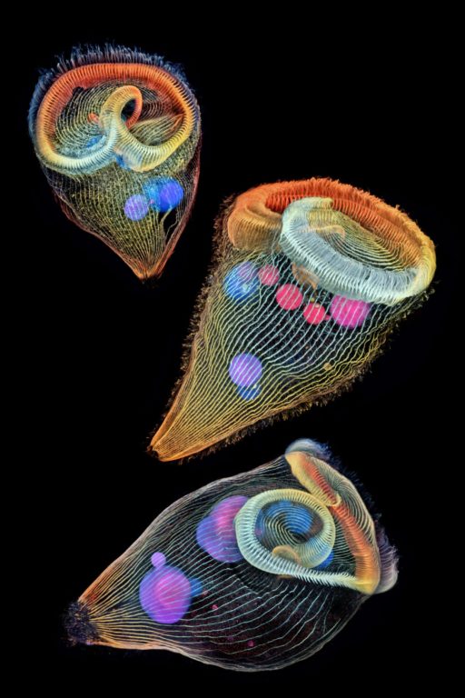
2nd Place: Depth-color coded projections of three stentors (single-cell freshwater protozoans) Photo credit: Dr. Igor Siwanowicz
In third place is Mr. Daniel Smith Paredes, who placed for his image of a developing American alligator embryo. He snapped this photo at around 20 days of development using immunofluorescence and is studying the development and evolution of vertebrate anatomy.
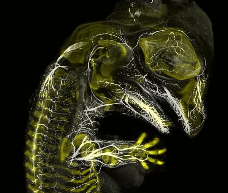
3rd Place: Alligator embryo developing nerves and skeleton
Daniel Smith Paredes Dr. Bhart-Anjan S. Bhullar

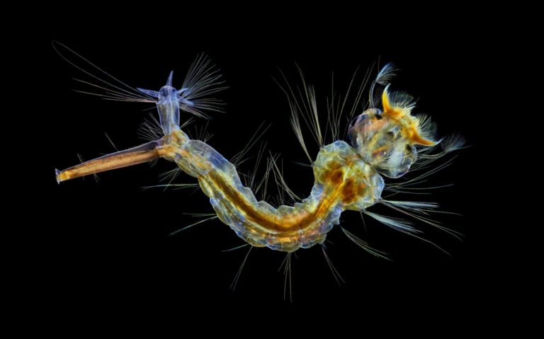
4th Place: Male mosquito. Photo credit: Jan Rosenboom
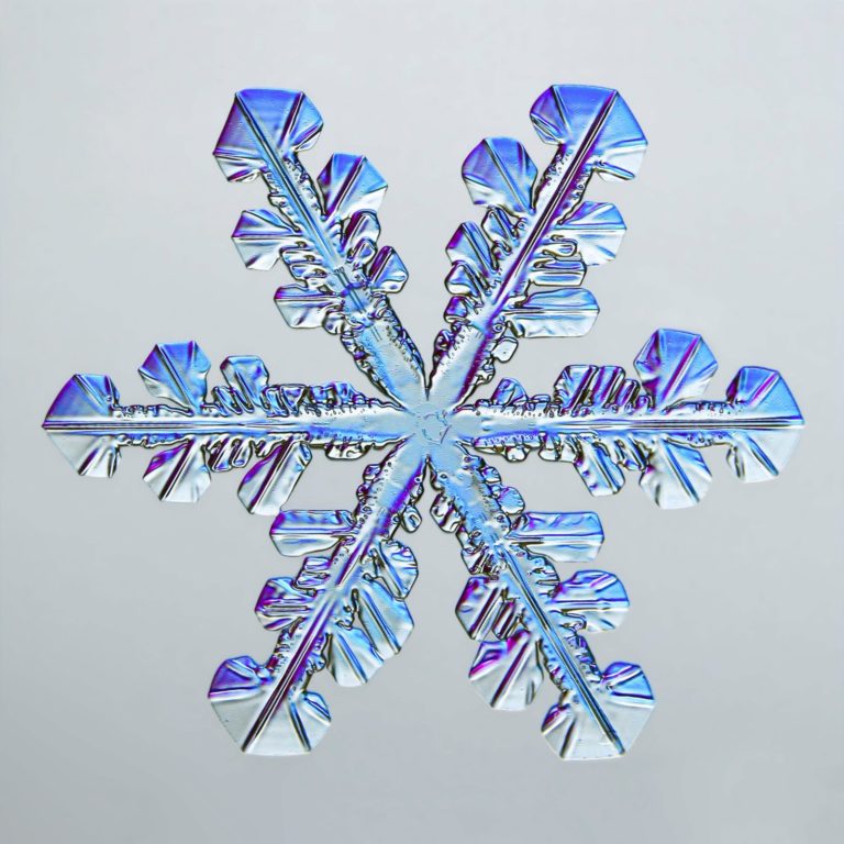
5th Place: Snowflake Photo credit: Caleb Foster

6th Place: Small white hair spider. Photo credit: Javier Rupérez

7th Place: Chinese red carnation stamen.
Photo credit: Dr. Guillermo López López

8th Place: Frozen water droplet. Photo credit: Garzon Christian

9th Place: Tulip bud cross section. Photo credit: Andrei Savitsky
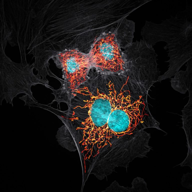
10th Place. BPAE cells in telophase stage of mitosis. Photo credit: Jason M. Kirk

11th Place: A pair of ovaries from an adult Drosophila female stained for F-actin (yellow) and nuclei (green); follicle cells are marked by GFP (magenta) Photo credit: Dr. Yujun Chen Dr. Jocelyn McDonald
12th Place: Mosquito larva Photo credit: Anne Algar

13th Place: Cuprite (mineral composed of copper oxide) Photo credit: Dr. Emilio Carabajal Márquez

14th Place: Female Oxyopes dumonti (lynx) spider Photo credit: Antoine Franck
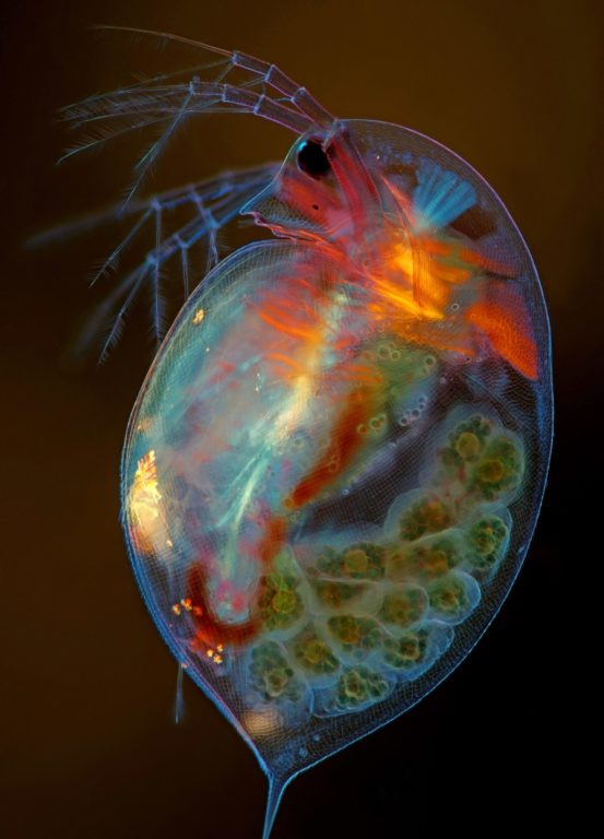
15th Place: Pregnant ‘Daphnia magna’ (small planktonic crustacean) Photo credit: Marek Miś
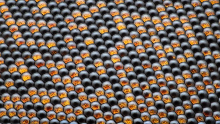
16th Place: A housefly’s compound eye. Photo credit: Dr. Razvan Cornel Constantin

17th Place Vitamin C. Photo credit: Karl Deckart

18th Place. Cristobalite crystal suspended in its quartz mineral host Photo credit: E. Billie Hughes
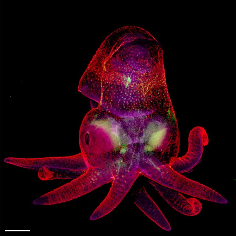
19th Place: ‘Octopus bimaculoides’ embryo Photo credit: Martyna Lukoseviciute Dr. Carrie Albertin

Blood vessels of a murine (mouse) heart following myocardial infarction (heart attack)
Photo credit: Simon Merz Lea Bornemann, Sebastian Korste
‘The Nikon Small World competition has been bringing stunning scientific images to the public for 45 years now,’ said Eric Flem, Communications Manager, Nikon Instruments, ‘Our goal has always been to show the world how art and science intersect. As new imaging and microscopy techniques develop over the years, our winners showcase these technology advances more and more creatively. First place this year is no exception.’
You may also like
Related Posts
Driving over the Berg River on Carinus Bridge, you’d probably dismiss Velddrif as just another...
read more
The tips of our fingers tell the story of who we are. Those faint undulating...
read more
Many parents across the world have opted out of traditional governmental and even private schooling...
read more